How Many Species of Bacteria are There? (with pictures)
Bacteria is a unicellular prokaryotic organism. The structure of the bacteria consists of three major parts: Outer layer (cell envelope), cell interior, and additional structures. Outer layer (Cell envelope): It includes the cell wall of bacteria and the plasma membrane beneath it.
Bacteria Cell Vector Art, Icons, and Graphics for Free Download
1. A bacterial cell remains surrounded by an outer layer or cell envelope, which consists of two components - a rigid cell wall and beneath it a cytoplasmic membrane or plasma membrane. ADVERTISEMENTS: 2.
Bacterial Structure Plantlet
Bacteria such as Clostridium difficile can become problematic and is now a serious type of nosocomial infection. The take home message is that [antibiotics increase selective pressure on bacteria which speed their evolution and create more unique and potentially pathogenic strains, for which our immune system cannot keep pace.].
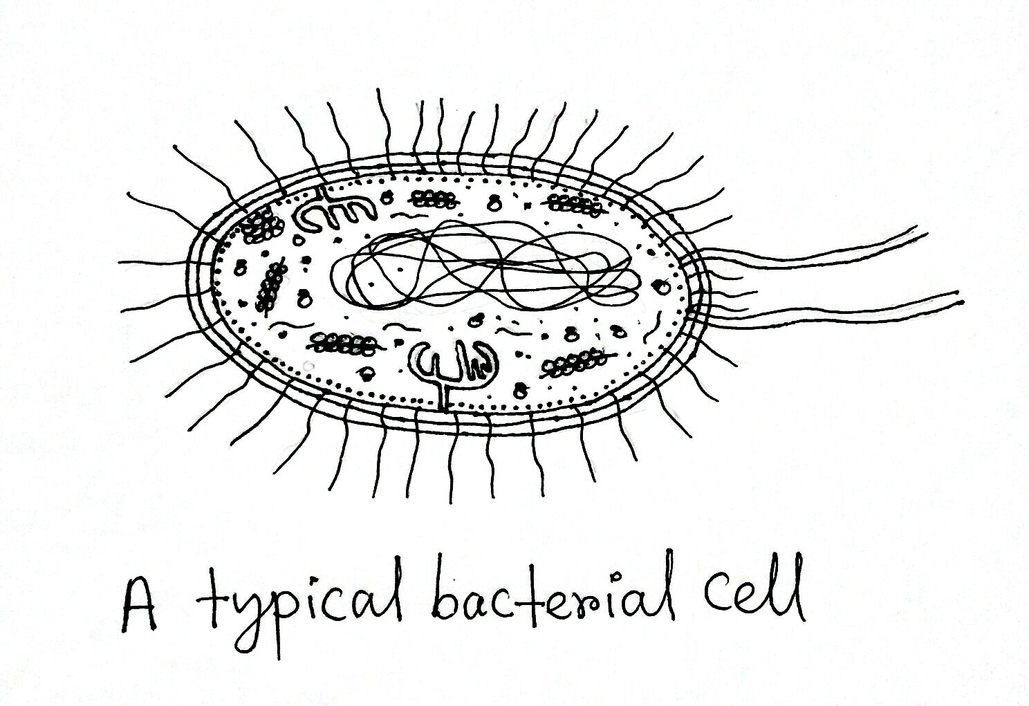
Structure and Function of a Typical Bacterial Cell with Diagram
Peptidoglycans are unique to prokaryotic organisms and consist of a glycan backbone of muramic acid and glucosamine (both N-acetylated), and peptide chains highly cross-linked with bridges in Gram-positive bacteria (e.g., Staphylococcus aureus ) or partially cross-linked in Gram-negative bacteria (e.g., Escherichia coli ).

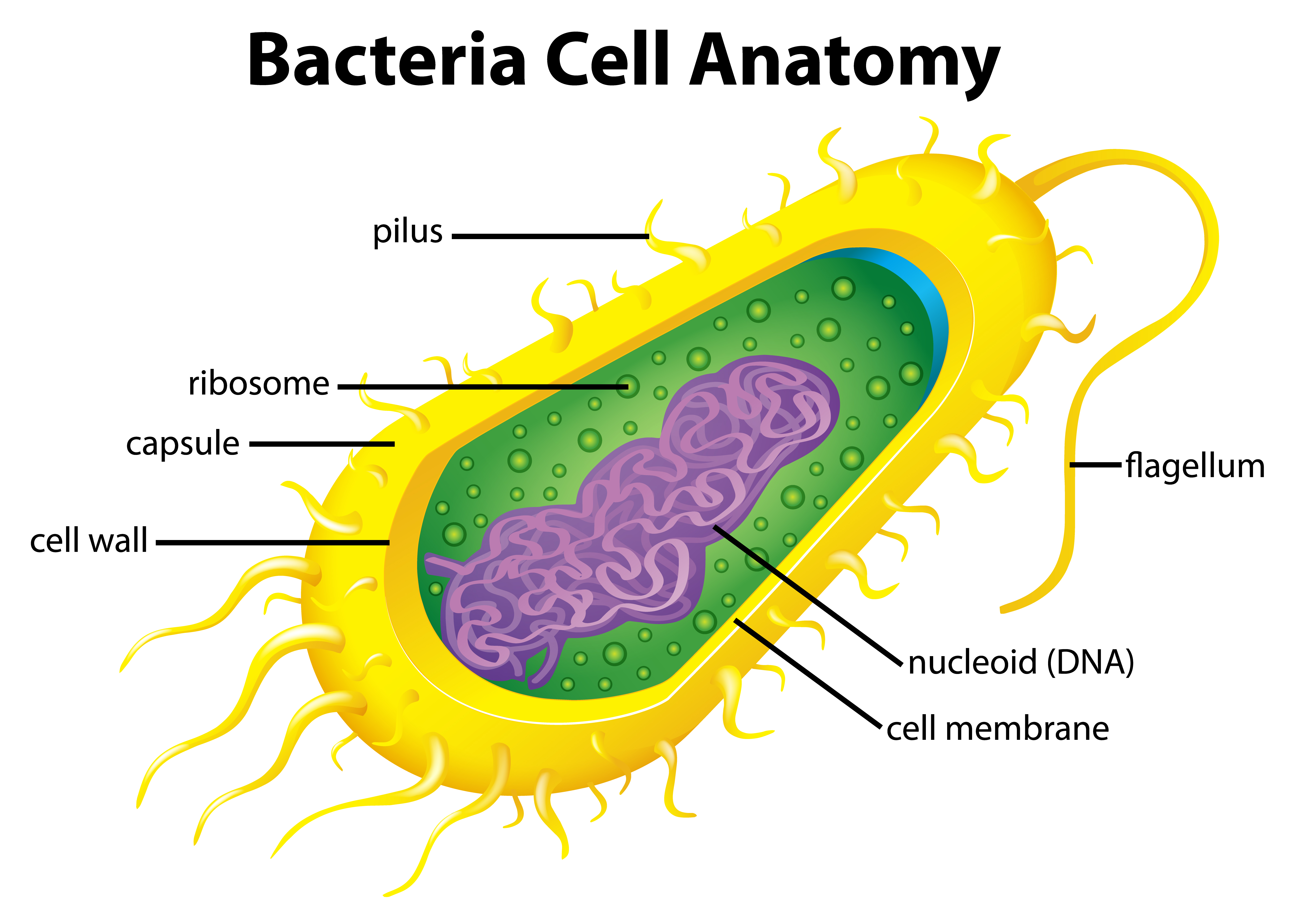
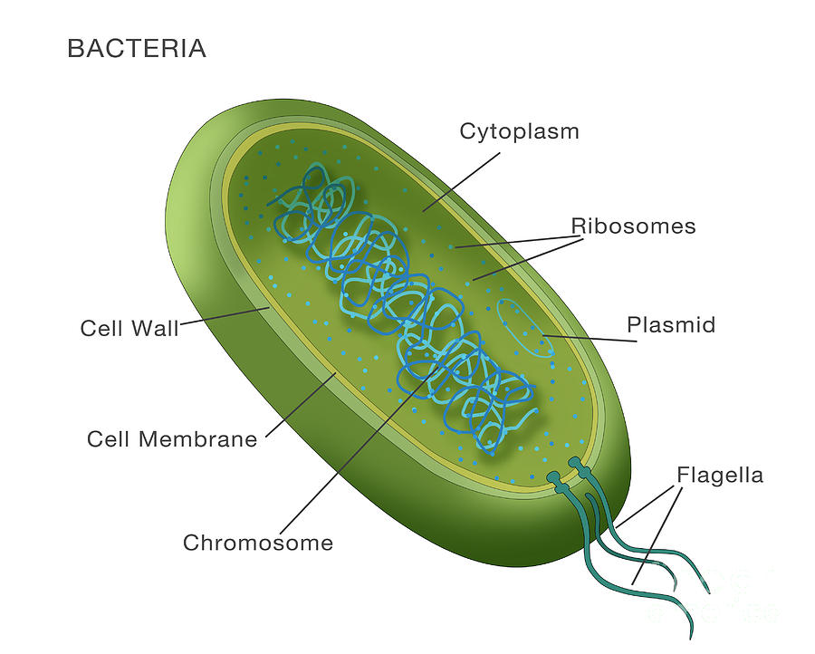
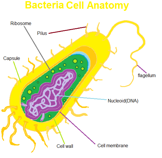
Bacteria cell anatomy Royalty Free Vector Image
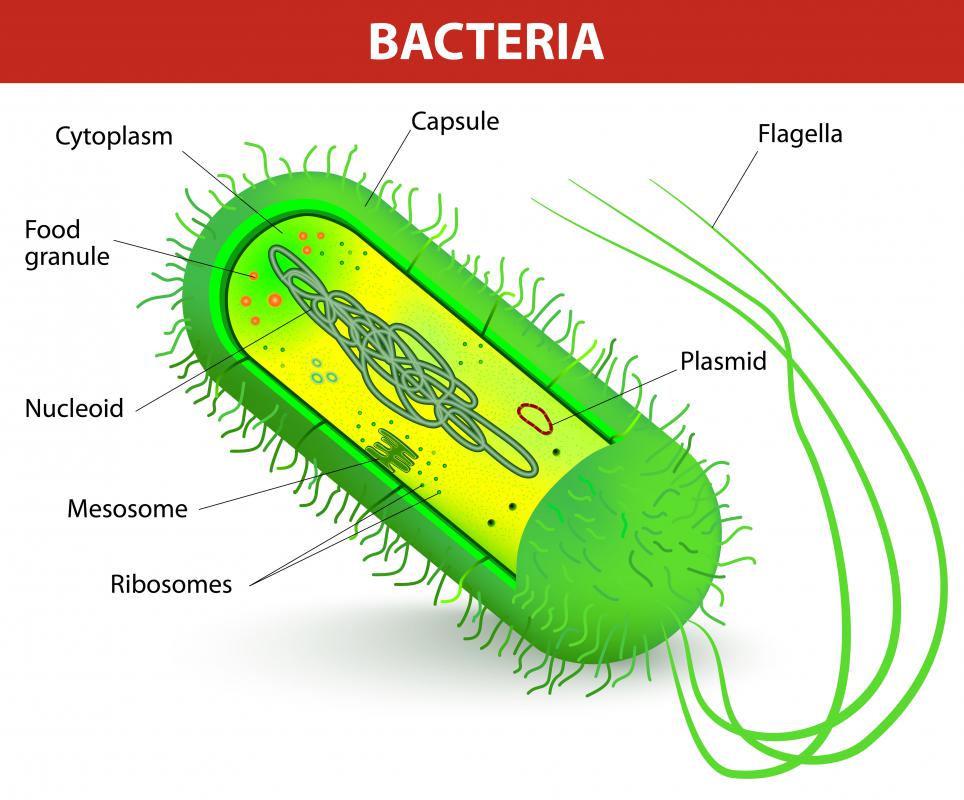
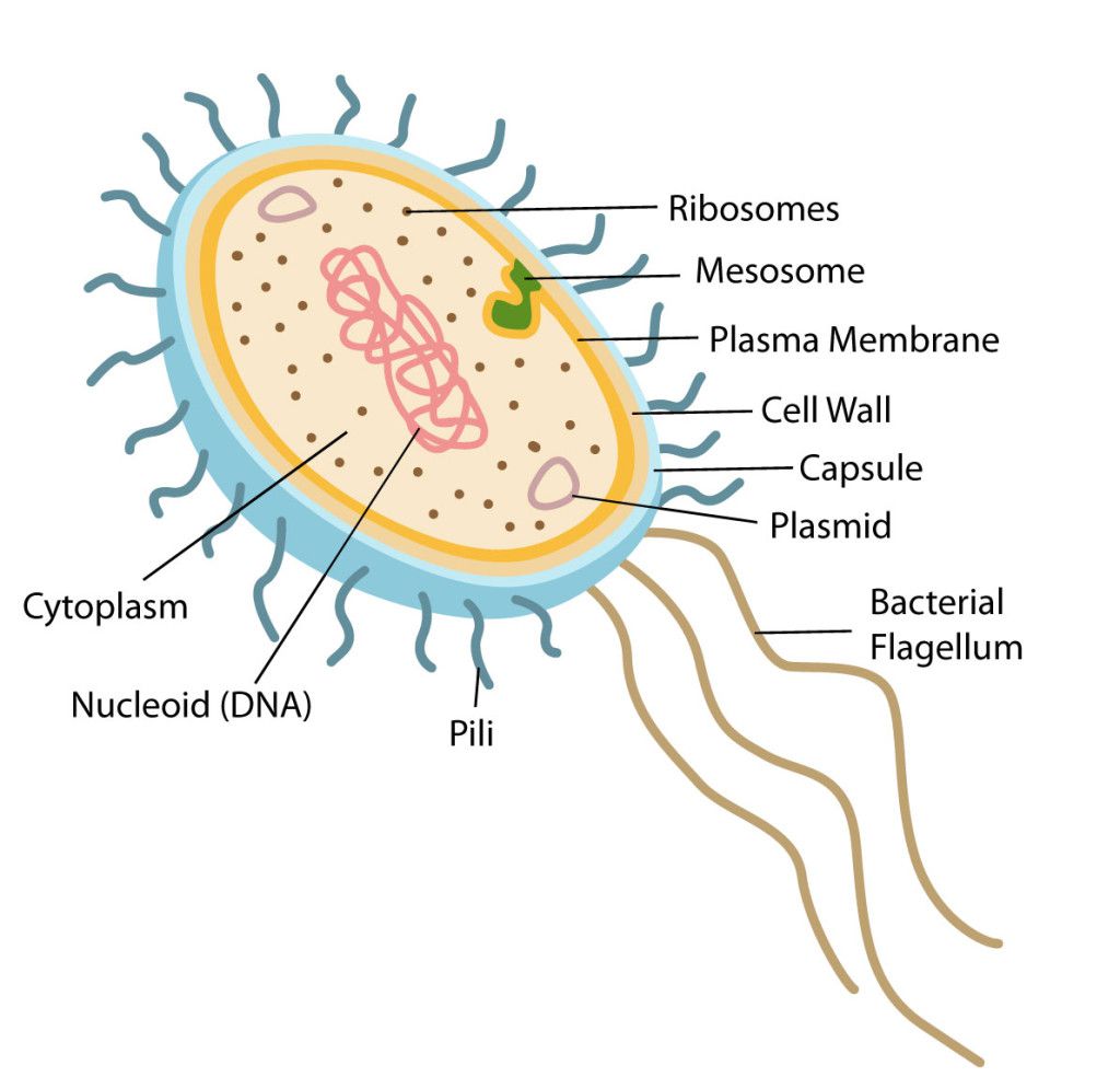
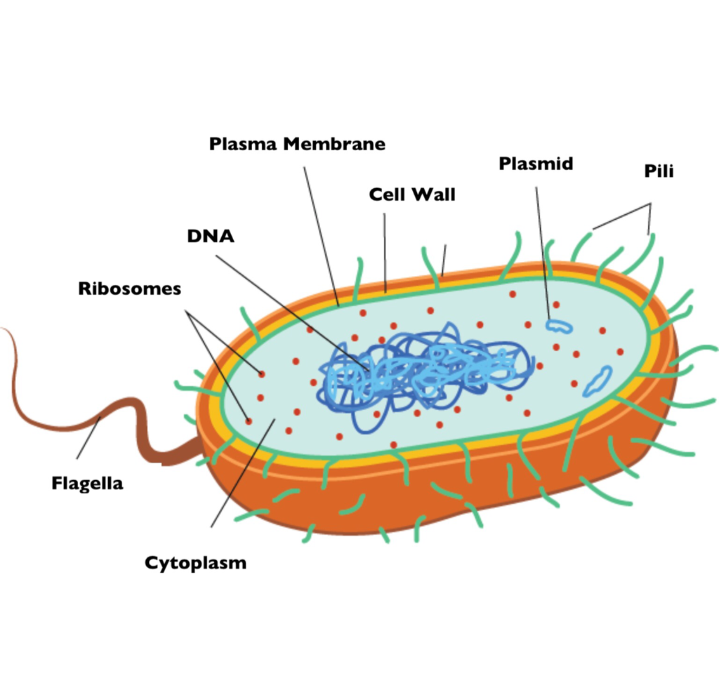
The bacteria diagram given below represents the structure of a typical bacterial cell with its different parts. The cell wall, plasmid, cytoplasm and flagella are clearly marked in the diagram. Bacteria Diagram representing the Structure of Bacteria Ultrastructure of a Bacteria Cell The structure of bacteria is known for its simple body design.
Bacterial Structure Plantlet
The three main shapes of bacteria are coccus, spiral, and bacillus. Cocci are bacteria that are spherical or ovoid in shape. Some cocci remain attached after binary fission, even though separate cells have been formed. For example, diplococci are cocci in pairs, streptococci are chains, and staphylococci are clusters of multiple cocci.
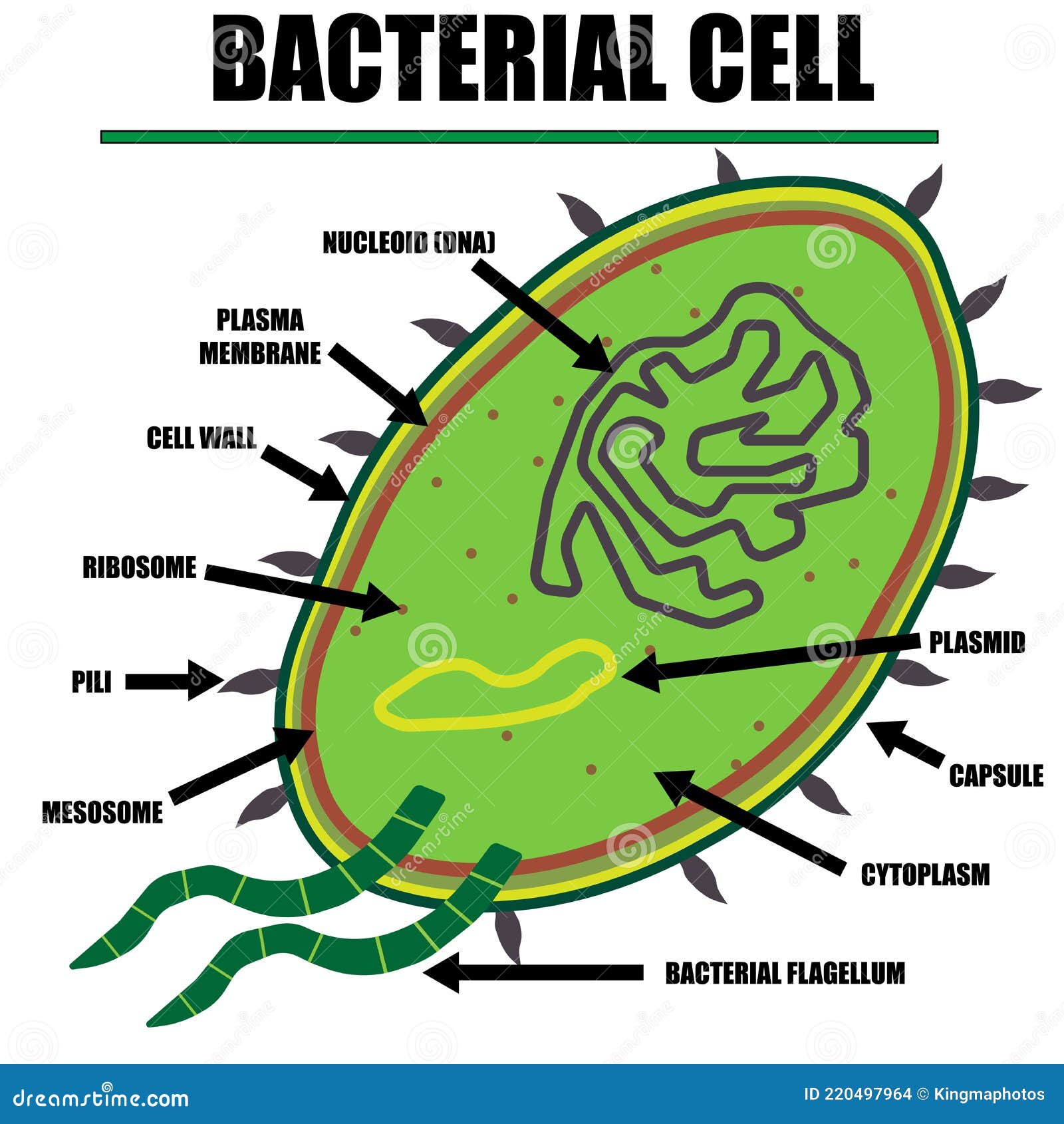
Bacterial Cell Color Diagram of Organelles Inside the Cell Wall for Science and Biology Concepts
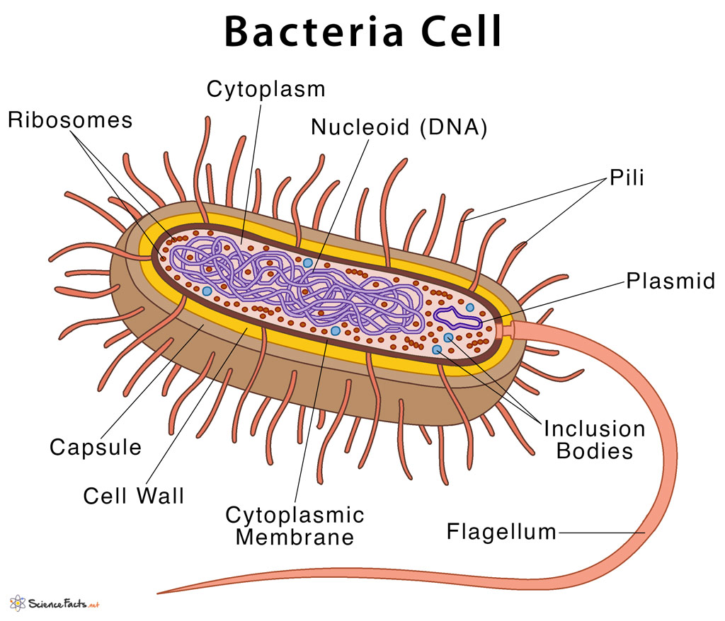
Bacteria: Definition & Characteristics With Examples & Diagram Bacteria Cell Bacteria are disease-causing, microscopic, single-celled organisms with prokaryotic cell structures. They do not have membrane-bound organelles, including a true nucleus.
Bacteria Diagram Photograph by Monica Schroeder
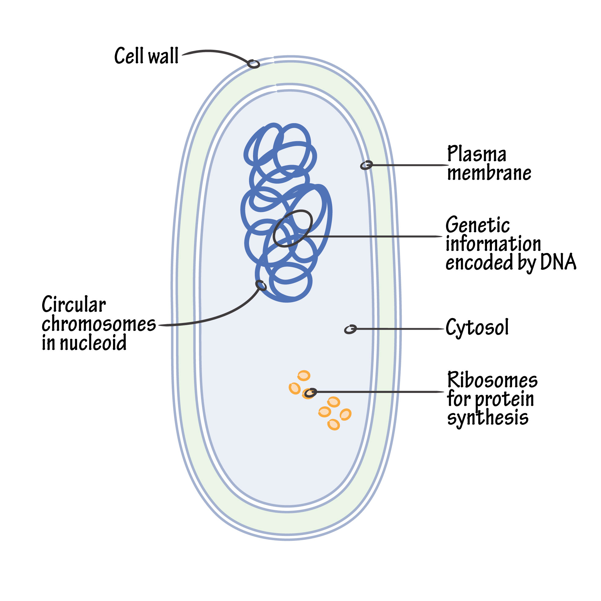
Diagram showing the relative sizes of some very small things including bacteria, which are typically around 1 to 2 μm in diameter (Source: Michigan Nanotechnology Institute for Medicine and Biological Sciences ). Image - Text Version Even though they are small, bacterial cells have many different parts.

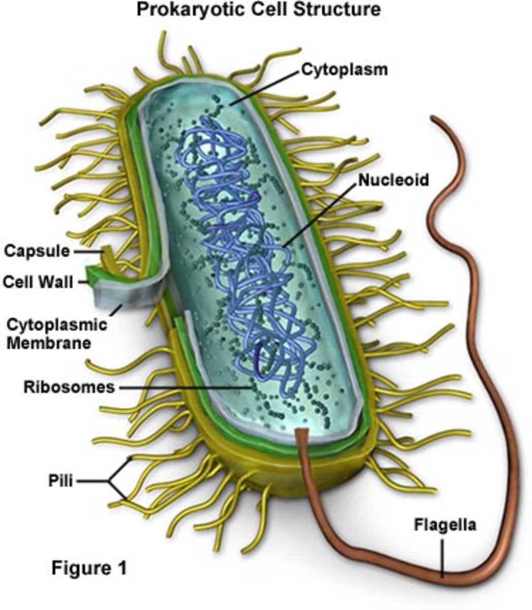
prokaryotic cell bacteria parts
Because of the simplicity of bacteria relative to larger organisms and the ease with which they can be manipulated experimentally, the cell structure of bacteria has been well studied, revealing many biochemical principles that have been subsequently applied to other organisms. Cell morphology [ edit] Bacteria come in a wide variety of shapes.
Bacterial Intracellular Structures That Give Bacteria/Prokaryotes an Advanatage! HubPages
Peptidoglycan (cell wall) Provides bacterial shape and rigidity. The cell wall consists of alternating units of N-acetylglucosamine and N-acetylmuramic acid. The polysaccharide chains are cross-linked by a peptide bridge. It is a primary target of antimicrobial therapy - because it is specific to prokaryotes.
Bacteria Grade 11 Biology Study Guide
What are prokaryotes? Prokaryotes are microscopic organisms belonging to the domains Bacteria and Archaea, which are two out of the three major domains of life. (Eukarya, the third, contains all eukaryotes, including animals, plants, and fungi.) Bacteria and archaea are single-celled, while most eukaryotes are multicellular.
Types and Structure of Bacteria ScienceAid
Bacteria - Definition, Structure, Diagram, Classification: Bacteria are truly fascinating microorganisms with an incredible ability to adapt and thrive in diverse environments. From their unique structures to their various nutritional and reproductive strategies, they play essential roles in shaping our world.

Bacteria Diagram
The structure of a typical bacterial cell with its various components is depicted in the bacteria diagram provided below. Table of Contents Bacteria Diagram with Labels Bacterial Cell Internal Structures Cell Wall Bacterial Capsule Pilus (plural Pili) Plasma membrane Cytoplasm Ribosomes Flagellum Nucleoid Plasmid Bacteria Diagram with Labels
Characteristics of bacterial cells
bacteria, any of a group of microscopic single-celled organisms that live in enormous numbers in almost every environment on Earth, from deep-sea vents to deep below Earth's surface to the digestive tracts of humans. Bacteria lack a membrane-bound nucleus and other internal structures and are therefore ranked among the unicellular life-forms.
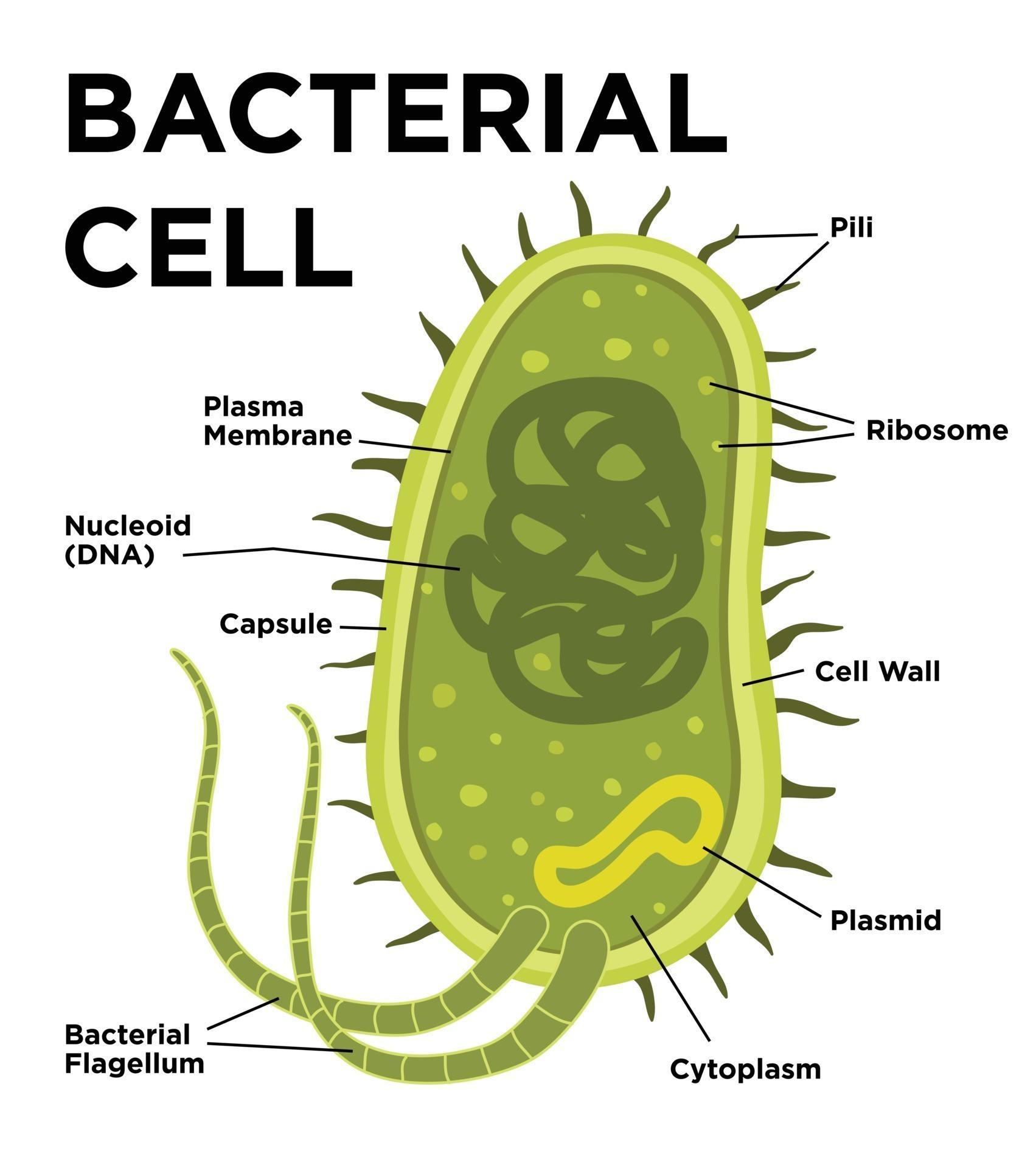
Bacterial cell anatomy in flat style. Vector modern illustration. Labeling structures on a
Bacterial DNA is fragmented and viral DNA is replicated. New viral particles are made and exit the cell. One contains host DNA instead of viral DNA. When this virus infects a new host, it injects the bacterial DNA, which can recombine with the chromosome of the new hows.. In this diagram, a transposon in the bacterial chromosome is copied.
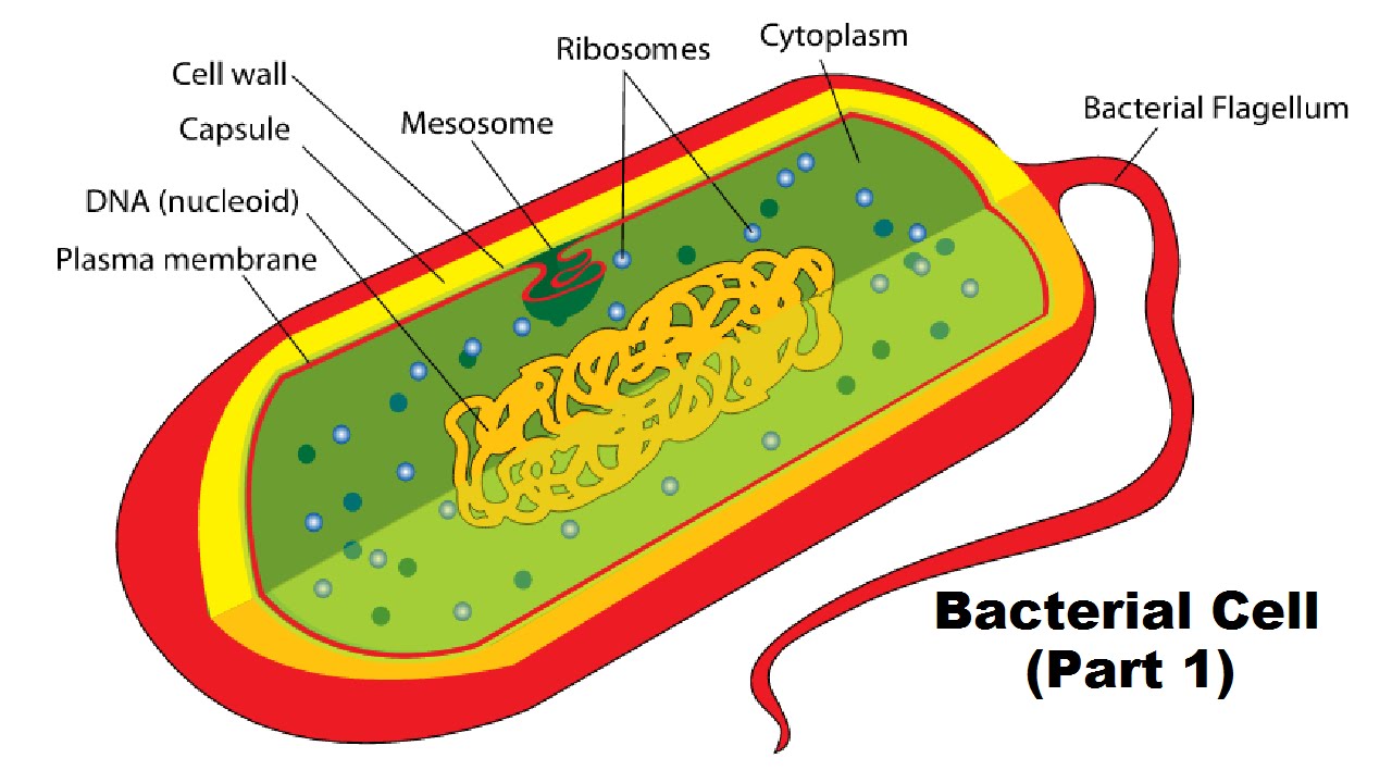
Structure of a Bacterial Cell (Part 1) YouTube
The nucleoid and some other frequently seen features of prokaryotes are shown in the diagram below of a cut-away of a rod-shaped bacterium. _Image credit: modified from "Prokaryotic cells: Figure 1" by OpenStax College, Biology, CC BY 3.0 _
